
Molecular Core Photos
To use this equipment, contact Dr. Grozdanov at molcore@ttuhsc.edu
or phone: +1 (806) 743-4624
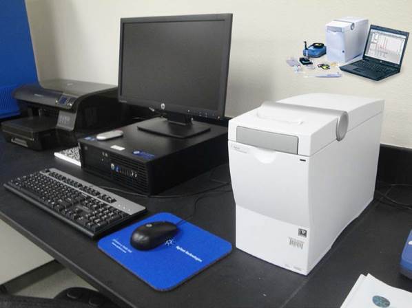 |
| 2100 Bioanalyzer (Agilent). The instrument uses microfluidics to resolve RNA, DNA, and protein samples. It is
useful to determine the quality of RNA (RNA integrity number) before running microarrays,
RNA-seq or real time quantitative PCR measurements. The individual samples are analyzed
and a pseudogel image is created. The nucleic acid or protein bands are sized and
quantified. The instrument requires minimal amount of sample (4µl). It replaces time-consuming
techniques associated with agarose gels or SDS-PAGE with fast, automated, high quality
digital data. Preparation time is 5 min and digital data is obtained in 30 minutes. More information about the instrument is available on the company's webpage. |
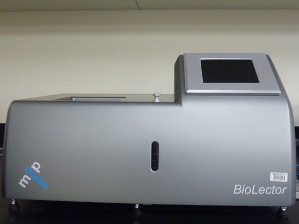 |
| BioLector (M2P labs). The instrument can be used to grow bacteria, yeast, or mammalian cells in suspension
in a 48-well plate. Cell growth (biomass), fluorescence, acid production (pH) and
oxygen consumption can be continuously and simultaneously monitored. CO2, oxygen,
humidity and temperature are also fully controlled and adjusted. The instrument allows
rapid optimization of growth parameters and implementation of cytotoxicity bioassays.
A companion for this system is a shaker (Multitron II; offline) that allows performing
parallelized experiments, thus freeing the time in the BioLector. More information about the instrument is available on the company's webpage. |
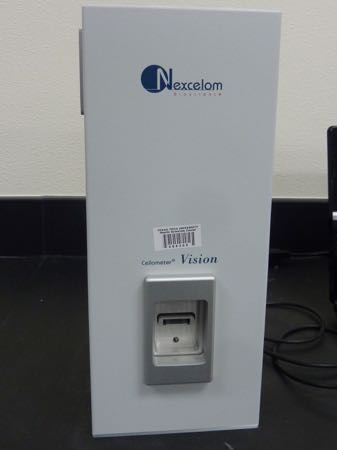 |
| Cellometer Vision Image Cytometry (Nexcelom Bioscience). The instrument can be used for simple image cytometry in a 20 µl sample, capable of
performing cell cycle analysis, apoptosis, autophagy, cell proliferation, mitochondrial
membrane potential, viability assays, morphology analysis and cell counting. Equipped
to monitor fluorescence of GFP, FITC, Acridine Orange + DNA, Acridine Orange + RNA,
RFP, PI, PE, as well as other commonly used fluorochromes in green and red spectra.
It captures images using an integrated digital camera and 20x objective. More information about the instrument can be obtained on the company's webpage. |
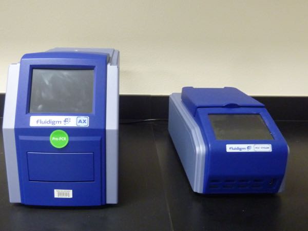 |
| Access Array System (Fluidigm). The access array is a high throughput target-enrichment system. It can enrich multiple
unique targets (e.g. exons) from a large number of samples, all at the same time.
Multiple samples and amplicons can be simultaneously barcoded (indexed), which streamlines
next generation sequencing library preparation. More information about the instrument is available on the company's webpage. |
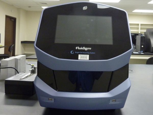 |
| C1 Single-Cell Auto Prep System (Fluidigm). Useful to study single-cell gene expression, single-cell mRNA analysis, and single-cell
genomics. It uses microfluidic technology that allows rapid and reliable isolation,
processing, and profiling of 96 individual cells. The use of C1 Single-Cell Auto Prep
System can be combined with the Access Array System (see above). More information about the C1 Single-Cell Auto Prep System is available on the company's webpage. |
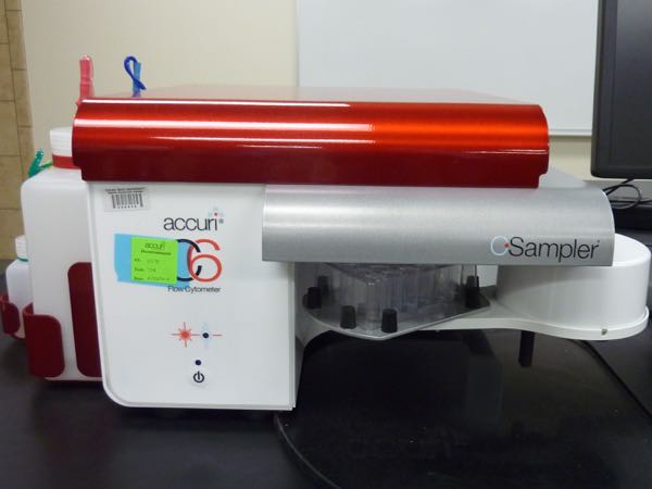 |
| BD Accuri C6 flow cytometer (BD Biosciences). The system is equipped with a blue and red laser, and four fluorescence detectors
with optical filters optimized for the detection of most common fluorophores (e.g.,
FITC, PE, PerCP, and APC). Any brand of 12 x 75 mm sample tubes and/ or microcentrifuge
tubes can be used for the sample preparation. The instrument is also capable of automated
data collection from 48- and 96-well plates. It can be used to study many cellular
functions, including apoptosis, cell cycle, cell proliferation, mitochondrial membrane
potential, immunoassays, cell phenotyping, cytosolic calcium and pH, nitric oxide. More information about the instrument is available on the company's webpage. |
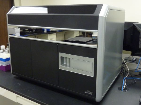 |
| ImageStream Mark II (Amnis). This system combines the speed, statistical power, and fluorescence sensitivity of
flow cytometry with the functional insights (e.g. morphology, fluorescent localization)
of high-resolution microscopy, making it a high-resolution microscopy in flow. It
is equipped with 20x, 40x and 60x objectives, 4 lasers (405 nm, 488 nm, 561 nm, 642
nm, 785 nm), and 12 imaging channels. Dual CCD cameras are used as detectors. This
system can be used to evaluate many different fluorochromes in non-adherent cells,
i.e., cells in suspension alive or fixed. More information about the ImageStream Mark II is available on the company's webpage. |
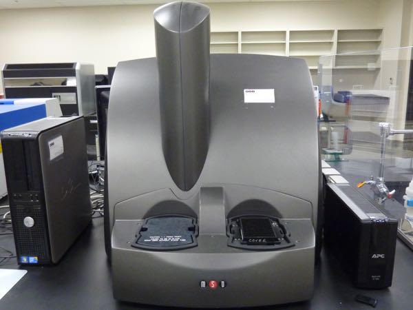 |
| SECTOR Imager 2400 (Meso Scale Discover, MSD). This multi-array technology (96- and 384-well, small spot) enables the detection of
biomarkers in single and multiplex formats. It utilizes special kits for profiling
biomarkers, cell signaling pathways, cancer markers, angiogenesis, cardiovascular
diseases, neurodegenerative disorders, apoptosis, metabolites, cytokines and others.
It can also be customized for individual applications. It is equipped with an ultra-low
noise CCD camera for high dynamic range, sensitivity and rapid read times. |
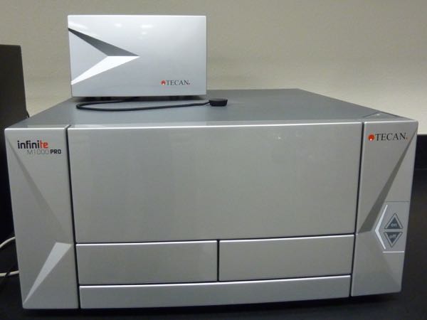 |
| Infinite M100 PRO Quadruple monochromator microplate reader (Tecan). Automated microplate reader for UV, VIS absorption, fluorescence intensity, and fluorescence
polarization with time resolved fluorescence, fluorescence resonance energy transfer
and alpha screen technology. It can also quantify luminescence and luminescence spectra.
Temperature controlled system (up to 42°C) and orbital and linear shaking capabilities.
Accommodates 6-, 12-, 24-, 48-, 96-, 384-, and 1536-well plate formats. The spectral
resolution is 1 nm. More information about the Infinite M100 PRO is available on the company's webpage. |
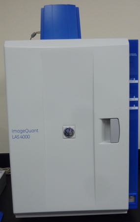 |
| ImageQuant LAS 4000 (GE Healthcare Life Sciences). The instrument is a multipurpose 16-bit 3.2 megapixel CCD camera system for sensitive
and quantitative imaging of chemiluminescence, standard UV transillumination, or white
light gel or blot documentation. More information about this instrument can be accessed on the company’s webpage. |
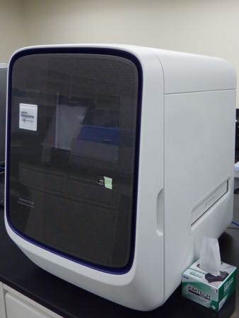 |
| Quant Studio 12K Flex real-time PCR system (Life Technologies). The instrument can be used for gene expression analysis, microRNA profiling, and non-coding
RNA analysis using TaqMan or SYBR Green assays. It supports real-time PCR in 96- and
384-well plate formats as well as TaqMan Low density Array cards. For more information click here. |
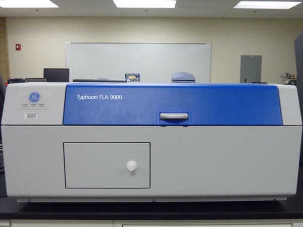 |
| The Typhoon FLA 9000 Biomolecular Imager is a variable mode laser scanner for flexible, sensitive imaging and quantitation
of proteins, nucleic acids, and other molecules using filmless autoradiography, fluorescence,
chemifluorescence, and gel documentation. For more information please click here. |