
Imaging Core Equipment Photos
To use this equipment, contact Dr. Grozdanov at imagecore@ttuhsc.edu or phone: +1 (806) 743-4624
 |
|
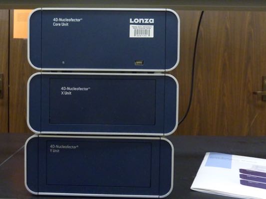
4D-Nucleofector Core X/Y (Lonza). The X unit supports nucleofection of various cell numbers in different formats and
it is used to transfect cells in suspension. The Y unit enables transfection of adherent
mammalian cells in 24-well plates. Cells can be transfected with either DNA or RNA.
Information about the 4D-Nucleofector is available on the company’s website. |
 |
|
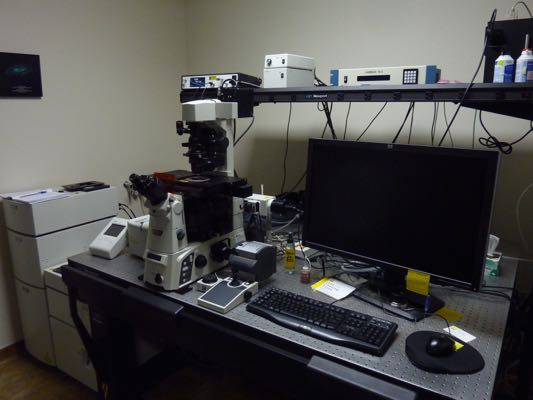
Nikon T1-E microscope with A1 confocal and STORM super-resolution modules. The system allows imaging of live or fixed cells and tissues labeled with fluorophores
using conventional or confocal microscopy. In addition, the system is equipped to
perform a super-resolution microscopy, specifically STochastic Optical Reconstruction
Microscopy (STORM). Super-resolution microscopy allows a precise localization of a
fluorophore, which is beyond the diffraction limit of light. Super-resolution microscopy
achieves resolution ten times greater than conventional optical microscopy. The system
is also equipped to perform spectral imaging experiments and other state-of-the-art
fluorescent optical approaches (TIRF, FRET, FRAP). A high temporal and spatial resolution
EMCCD camera is attached to the system. The system is equipped with a stage top environmental
chamber for prolonged observation of living cells. |
 |
|
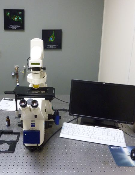
Zeiss Axiovert 200 M microscope. This microscope is one of the most popular microscopes in the Image Analysis Core
Facility. The microscope is inverted, fully motorized and equipped with 10x, 20x and
63x objectives for a wide filed fluorescence microscopy. Color and monochrome Axiocam
CCD cameras are attached to the microscope, allowing capture of colored samples (e.g.
H&E, peroxidase stained samples) or low-intensity florescent samples (e.g. YFP, GFP,
RFP and DAPI), respectively. The objectives and filters also permit acquisition of
phase contrast and differential interference contrast (DIC) images. The microscope
stage is equipped with a holder that accommodates various sizes cell culture dishes
and plates, allowing a quick examination of transfection efficiency. |
 |
|
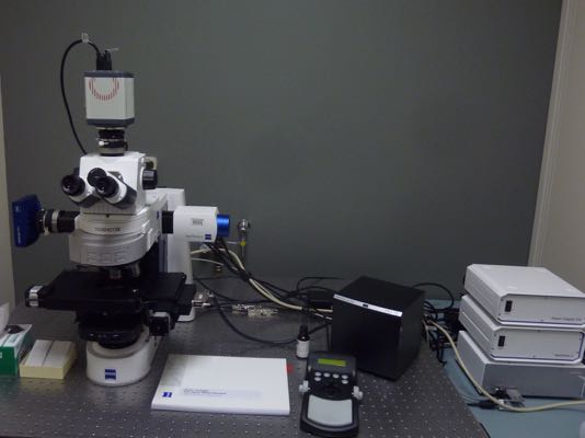
Zeiss Axio Imager M2 microscope for stereoscopic analysis. This system is fully motorized and configured for bright field and multi-channel fluorescent
imaging. The microscope is equipped with x, y, z motorized stage and Zeiss Apotome
system to create focal optical sections through your thick specimens using structured
illumination. Images are acquired with a low noise CMOS chip 2048 x 2048 pixels monochrome
digital camera (ORCA-Flash4.0, Hamamatsu). It works with Stereo Investigator, Neurolucida,
and deconvolution software (MBF Bioscience). |
 |
|
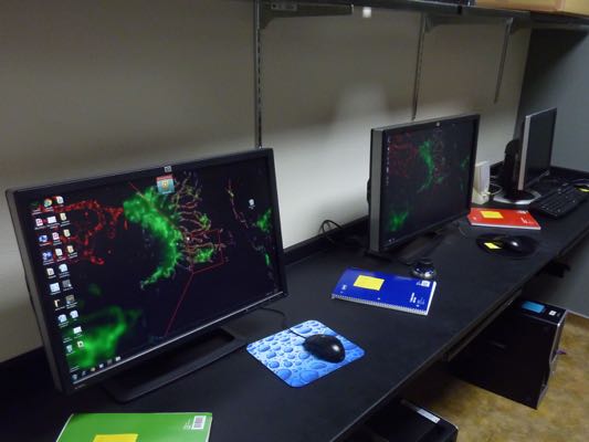
Workstation for data analysis. Several computers are available for off-line analysis of digital images created in the facility with all necessary software for image data analysis. |